
Serendip is an independent site partnering with faculty at multiple colleges and universities around the world. Happy exploring!
The Rat Forebrain
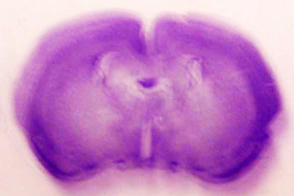
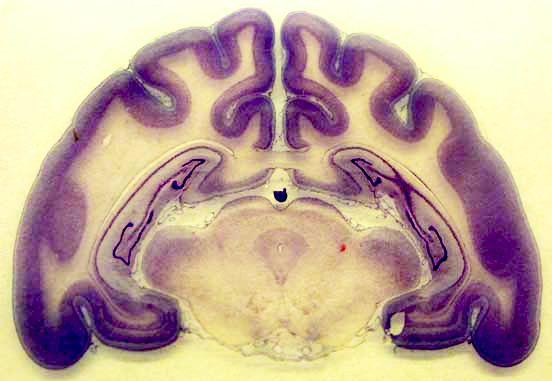
Now we are looking at a cross-section of the forebrain of a rat.
As with the human, monkey, and cat forebrains, this image is a good illustration of the cerebral cortex at the frontal lobe of the brain. The dark purple that looks like an outline is gray matter. The light purple inside the dark purple is white matter. You can also see the body of the corpus callosum, which is the structure connecting the two hemispheres of the brain. The white elliptical openings within the brain are ventricles, which are fluid-filled cavities that serve to cushion and protect the brain. You can also see midbrain structures in this image. What else do you notice about this image?
Does this look different from what you saw in the human, monkey, and cat forebrains? What is similar? What is different? Compare this with the human forebrain. Or compare with the monkey forebrain. Compare this with the cat forebrain, too. Do you notice any differences in amounts of or degree of cortical folding?
Where shall we go from here? Click on your browser's "back" button to return to the midsagittal view of the rat brain, or click on one of the buttons below to learn about these structures, to slice other brains, or to move on towards magnification.


Remote Ready Biology Learning Activities
Remote Ready Biology Learning Activities has 50 remote-ready activities, which work for either your classroom or remote teaching.
What's New? Subscribe to Serendip Studio
A Random Walk
New Topics
-
11 weeks 13 hours ago
-
11 weeks 1 day ago
-
11 weeks 4 days ago
-
11 weeks 4 days ago
-
11 weeks 4 days ago

Comments
Post new comment