
Serendip is an independent site partnering with faculty at multiple colleges and universities around the world. Happy exploring!
What's the Point?
After looking at all these sections and slides and close-ups of the various types of brains, what have we learned? What kinds of trends have we observed? What are we still curious about?
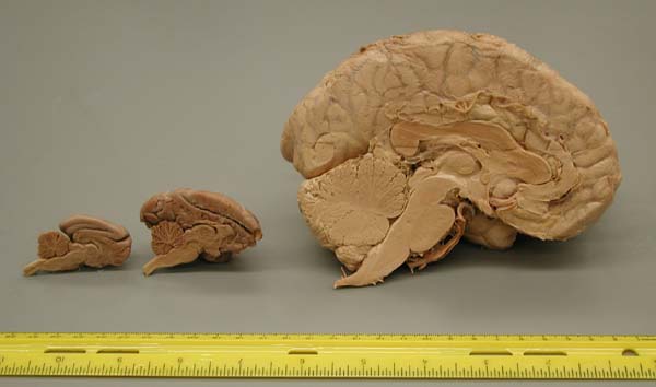
When you saw the sagittal slices of all the brains, what did you notice? Comparing across the sections you probably noticed that there were some general differences between the brains (involving size, shapes of structures, etc). For example, it may seem easier to differentiate structures in the human brain as opposed to the rat brain. It has been found that as one ascends a phylogenetic sequence, there is clearer definition and demarcation of structures, almost as if the brain is better organized. Yet, the closer we got, especially when we made forebrain and hindbrain slices, the more you may have realized that the brains and their structures look quite similar. In other words, yes, there are differences between animals' brains but also deep and fundamental similarities. Here are some pictures to remind you:
 |
 |
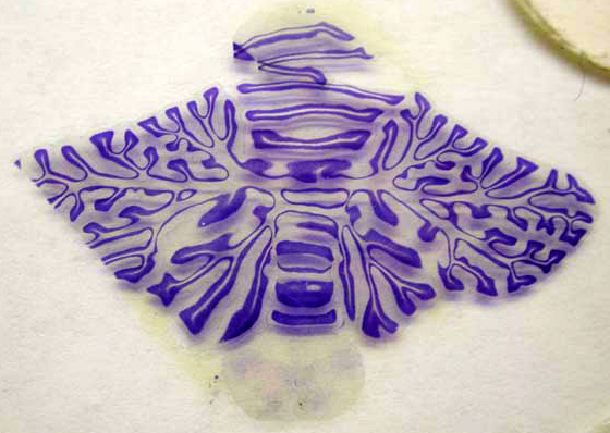 |
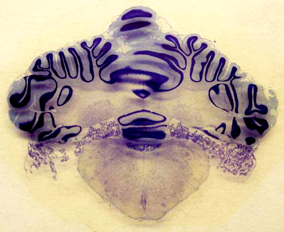
| Cat Hindbrain | Cat, Monkey, Human -- Midsagittal View | Monkey Hindbrain |
As you can see above, both the cat and monkey cerebellums look rather similar. The structure is located in similar places on each respective brain, above the brainstem, and is rather convuluted. Yet, there are also general differences that make it easy to differentiate one from another.
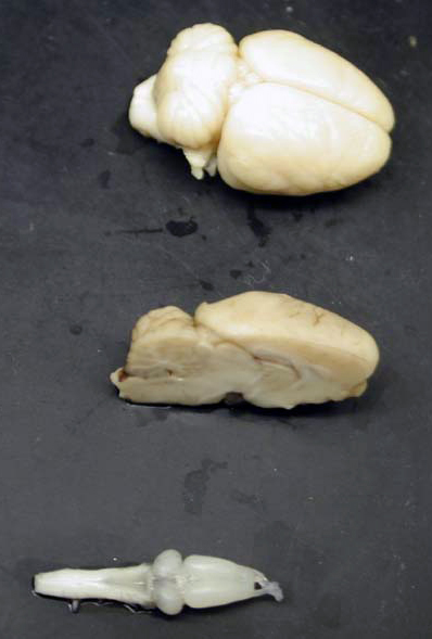
Another interesting comparison is between the rat and frog brains. Look at the picture to the right and note any similarities and differences. This may be difficult for you to see due to the fuzziness of the photograph, but stick with me. The frog brain is on the bottom; the middle brain is a sagittal view of the rat brain; the top brain is a full view of a rat brain. Essentially, the frog brain resembles the basic structures of the rat brain, laid out flat. For example, you see the spinal cord leading to the brain stem and cerebellum and then the midbrain/forebrain structures. So, in this way, the rat and frog brains are quite similar. Yet, in the rat brain we find neocortex surrounding the mid- and forebrain, which we cannot see in a frog brain. Indeed, frogs do not have neocortex (remember -- only mammals have this multi-layered structure). Click here for a discussion about this.
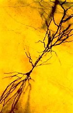 |
What if we look even closer, at the cellular level? What if we magnify these pictures so that we can see individual neurons? |

Remote Ready Biology Learning Activities

Remote Ready Biology Learning Activities has 50 remote-ready activities, which work for either your classroom or remote teaching.
What's New? Subscribe to Serendip Studio
A Random Walk
New Topics
-
9 weeks 6 days ago
-
10 weeks 6 hours ago
-
10 weeks 3 days ago
-
10 weeks 3 days ago
-
10 weeks 3 days ago

Comments
Fyre
Fyre’s Monologue:This is the character of Fyre who wants the tree where apples are lying.
Post new comment