
Serendip is an independent site partnering with faculty at multiple colleges and universities around the world. Happy exploring!
Brain imaging: similarities and differences between individual brains
Imaging Similarities and Differences Among Brains
created for Biology 202 at Bryn Mawr College
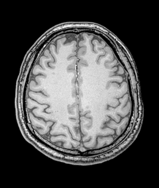
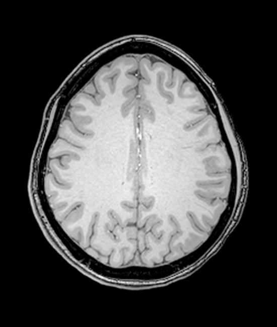
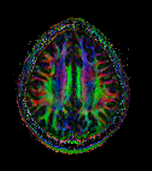
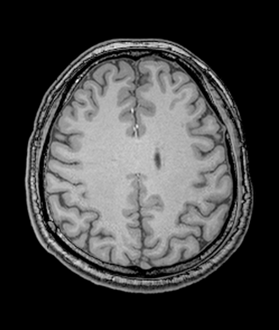
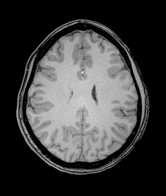
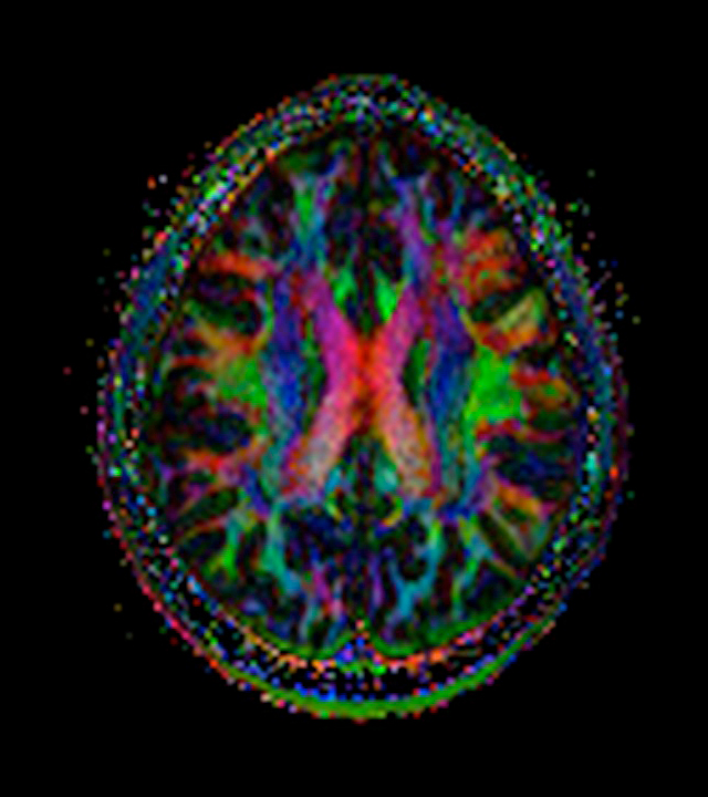
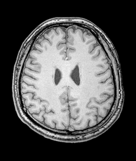
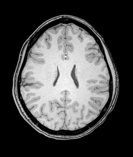
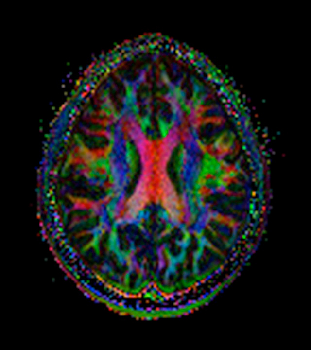
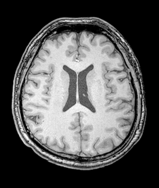
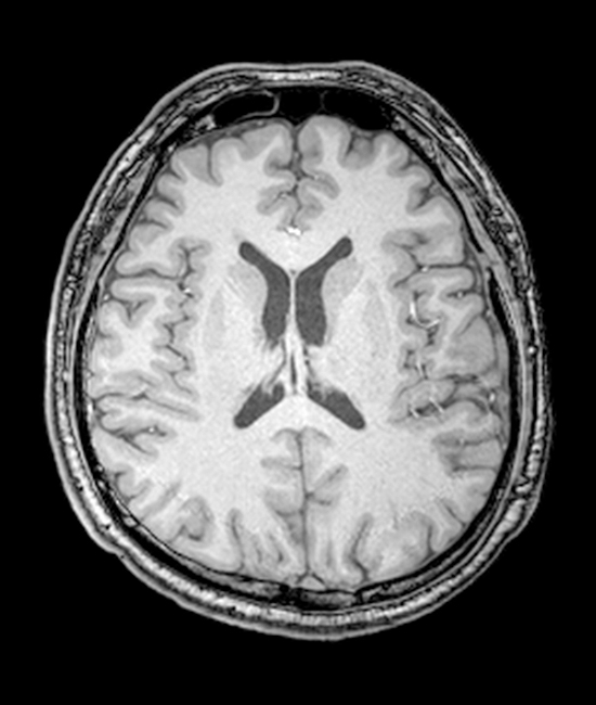
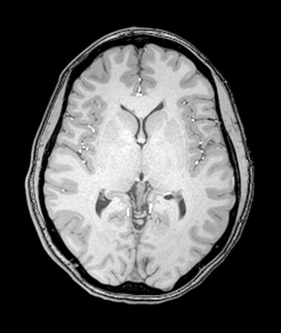
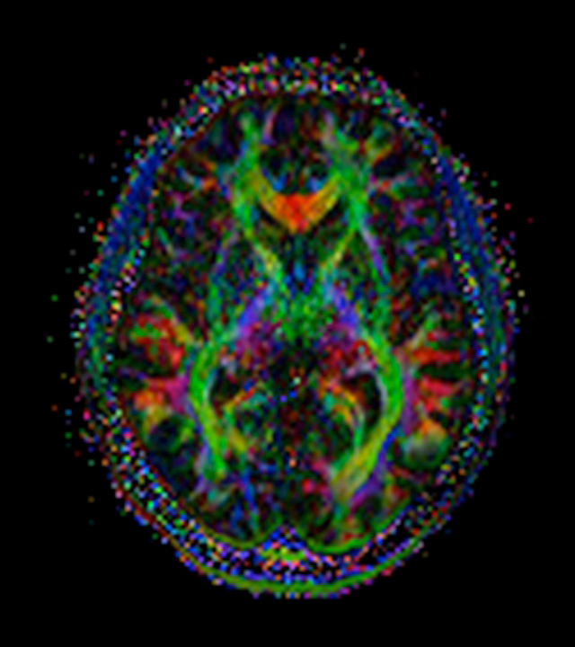
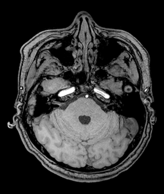
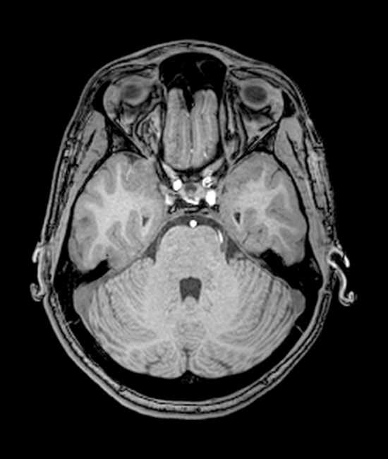
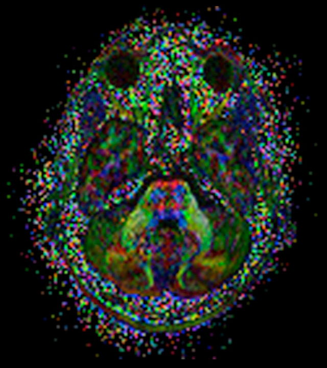
The past twenty years has seen great advances in the development of new technologies to visualize the brain, and the pace is increasing. Shown below are examples of magnetic resonance imaging (MRI) and diffusion tensor imaging (DTI). As the ability to image the brain becomes more sophisticated, so too does the evidence that the brains of individual humans are "sort of the same but sort of different." The sets of MRI images are comparable sections of the brains of two different individuals (left two MRIs of different individuals, color images are of individual #2).
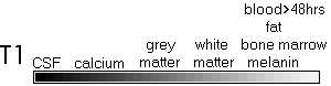
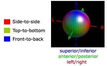
T1 MR1 (first and second images below) yields a grayscale image from which one can differentiate different tissue types. In T1 images, white matter appears white (axonal fiber bundles), gray matter appears gray (the cortex), and cerebral spinal fluid (within the ventricles) appears the darkest. Color maps are one type of image that can be generated from DTI data (third image). Color corresponds to the direction in which white matter bundles run. Red fibers run from side-to-side (e.g., corpus callosum); blue fibers run superior/inferior (e.g., internal capsule); green fibers run rostral/caudal (e.g. infeior longitudinal fasciculus). The slices shown side-by-side below were chosen based on the anatomical landmarks described beneath each pair.
Top of the brain; Most ventral slice showing separation of hemispheres
Slice with the maximum anterior-to-posterior width of the corpus callosum
Most dorsal slice showing both lateral ventricles
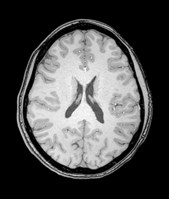
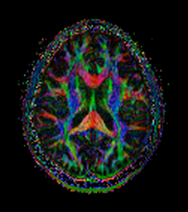
Slice in which lateral ventricles are the most closely approximated
Slice showing the insular cortex
Slice with the maximum width of the aqueduct
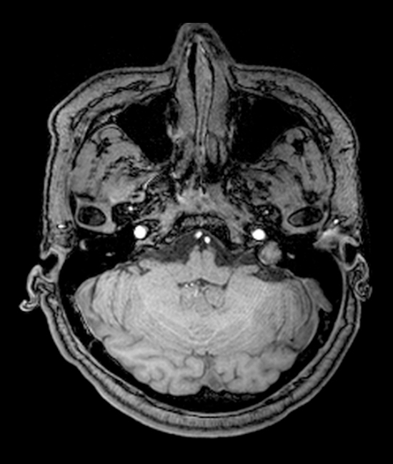
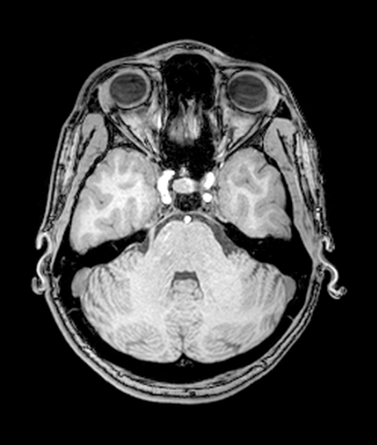
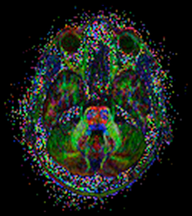
Slice with the maximum width of the cerebellum
Posted by Laura Cyckowski and Paul Grobstein 8 Feburary, 2010.
Remote Ready Biology Learning Activities
Remote Ready Biology Learning Activities has 50 remote-ready activities, which work for either your classroom or remote teaching.
A Random Walk
New Topics
-
9 weeks 6 days ago
-
10 weeks 8 hours ago
-
10 weeks 3 days ago
-
10 weeks 3 days ago
-
10 weeks 3 days ago

Comments
Post new comment