|
The exercise on functional neuroanatomy
you are about to complete was devised by five
students in the bi-college (Haverford and Bryn Mawr)
community: Jessica Kuhn, Jessica Magid, Karen Revere,
Elizabeth Caris, and Gray Vargas. The students
are enrolled in a Psychology/Biology seminar class called
“Topics in Neural and Behavioral Science”.
All of the students in the class devised lab exercises,
this is the one that was chosen. Several other students
from the class have assisted in preparing the lab for
you today and will be assisting you as you complete
the lab exercises.
The overall objectives of this
“Friday in the Lab” session are as follows:
-
To be introduced to the anatomy of the brain.
-
To explore the varied distribution of sensory
receptors in different parts of the body.
-
To study the functions of some of the sensory
systems.
-
To investigate the communication between the
two cerebral hemispheres.
In order to understand the directions
that follow in this lab manual, please refer to the
following background information:
Definitions:
-
Neuron – cells found in the brain
and body that transmit information between the
brain and the rest of the body
-
Central Nervous System: neurons (and their supporting
cells) found in the brain and spinal cord (encased
by bone)
-
Peripheral Nervous System: all other neurons.
-
Neurotransmitter – chemicals produced
by neurons that are released to carry their signal
to other neurons; they can excite or inhibit other
neurons
-
Axon – the extension of a neuron
that sends outgoing signals to the next neuron;
each neuron has one
-
Dendrite – the extensions of a neuron
that receives information from other neurons
-
Synapse – the space between neurons
where a signal is transmitted from one neuron
to another
-
Action potential – an electrical
signal that passes down the axon and stimulates
the release of neurotransmitters into the synapse
to transmit information to the next neuron
-
White matter – nervous tissue in
the brain and spinal cord that contain axons covered
in insulating material (myelin), rather than cell
bodies. Groups of white matter in the PNS are
called nerves groups of white matter in
the CNS are known as tracts; both are bundles
of axons located together
-
Grey matter – nervous tissue that
contains mostly cell bodies and axons and dendrites
that are not covered in myelin
-
Spinal cord – the column of nervous
tissue that runs from the brain down the back
of the body; it contains all of the “ascending”
tracts that carry information gathered from the
nerves in the skin, joints and muscles to the
brain. Also contains “descending”
tracts that carry information from the brain to
the nerves that leave the spinal cord to control
the muscles (produce body movements).
-
Cerebrum – the largest and most
developed part of the brain in humans that controls
most higher cognitive functions and voluntary
movements
-
Cerebral hemisphere – one half of
the cerebrum; each hemisphere has corresponding
structures, but some functions are controlled
more by one hemisphere or the other
-
Medulla – the part of the brain
stem that connect the brain to the spinal cord;
it controls involuntary functions such as breathing,
heart rhythms, and swallowing
-
Thalamus – the part of the brain
that receives and processes sound information
and transmits it to other parts of the brain
Sheep Brain Dissection – Gross NeuroanatomyEquipment:
- Sheep brains (2 per group)
- Dissecting knife
Objectives:
- To observe the overall organization of the brain
first hand.
- To prepare for the other stations by gaining an
understanding of where each of the relevant regions
is located.
Introduction:
We will be dissecting
a sheep’s brain as an example of a mammalian brain
that is relatively similar in organization to the human
brain. Since individual neurons are too small to be
seen by the naked eye, we will instead be examining
various regions of the brain to get an idea of what
function they perform.
Procedure: (if you do
not want to participate in the actual dissection you
can visit http://academic.uofs.edu/department/psych/sheep/framerow.html
and complete a virtual dissection)
-
Start by removing the meninges,
which are the 3 outer layers that protect the brain
and spinal cord, most of the outermost layer was
probably removed before you received the brain.
- Dura mater – outermost
membrane; tough and virtually opaque
- Arachnoid – middle
membrane; somewhat transparent
- Pia mater – innermost
layer; extremely delicate
-
If you look at the cerebrum, you
will notice that it is not a flat surface, but it
consists of sulci (grooves) and
gyri (ridges), which allow for
a large amount of material to be condensed into
a smaller amount of space. Look for the “T”
that is formed between the sulcus running down the
center of the cerebrum, and the deep groove (known
as the cruciate fissure) that runs
across the front third of the brain.
- The gyrus located after the cruciate fissure
is known as the primary somatosensory
cortex, which is the region where most
“touch” (including pressure, pain,
temperature, etc.) information is received from
the periphery. The organization of this region
will be discussed at one of the stations.
-
Next, turn the brain over and locate
the cranial nerves. These nerves
are located on the bottom surface of the brain,
and they are arranged in pairs. Each pair of nerves
controls a different function associated with the
senses and the muscles of the face. For the purpose
of this lab you should try to locate the following
cranial nerves:
- Olfactory bulbs (part of the Cranial
Nerve I system: Olfactory nerve) –
two flaps of tissue located near the front of
the brain. Smell information is actually sent
to the olfactory bulbs through cranial nerve
I, but this was removed when the brain was removed
from the skull.
- Optic Chiasm (part of the Cranial
Nerve II system: Optic nerve) –
the structure behind the olfactory bulbs that
is shaped like an X. Since information about
each side of the visual field is received from
both eyes, the optic chiasm is the point where
the information from the two eyes is separated
to be sent separately to the two halves of the
thalamus, and then to the two hemispheres.
-
Take one of the 2 brains and cut
down the middle between the hemispheres. Now that
you are looking at the inside of the midline of
the brain try to locate a bundle of white fibers
that connects the two hemispheres. This bundle of
fibers is known as the corpus callosum,
and it transfers information between the cerebral
hemispheres.
-
Now you have some time to slice
the brains in a few different directions and to
look at the overall organization. The plates pictured
below are photographs of the sheep brain. They are
provided below so that you can try to locate the
different neural structures, and students and teachers
are available to help you identify the structures
and to tell you a little bit about their functions.
The following plates were downloaded
from the Department of Psychology at the University
of Guelph (Canada), where a sheep brain dissection manual
is maintained (http://www.psydev.uoguelph.ca/faculty/peters/labmanual/)
Figure 1. the ventral surface
of the brain

Figure 2. the midsagittal section
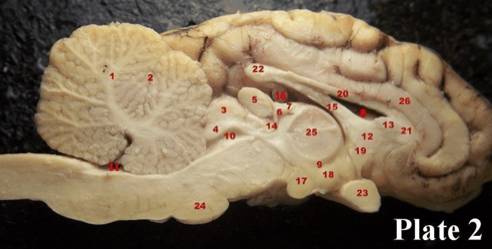
Figure 3. an anterior coronal
section
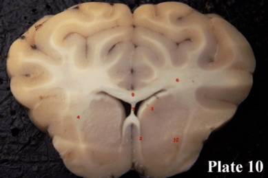
Figure 4. a posterior coronal
section
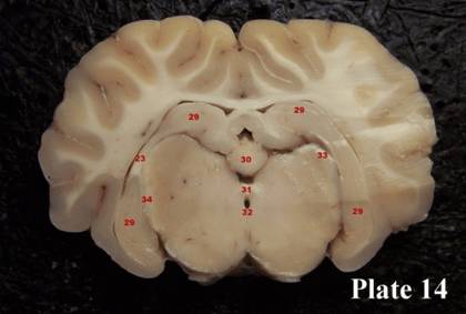
Table 1. Brain structures as
labeled in the plates above
Plate
1 Structures:
1 frontal cortex
2 olfactory bulbs
3 periamygdaloid cortex
4 optic chiasm
5 lateral olfactory tract
6 medial olfactory tract
7 pituitary gland (not seen in Plate 1)
8 mammillary bodies
9 cerebral peduncles
10 pons
11 trapezoid body
12 pyramidal tract
13 olive
14 olfactory nerve (not seen in picture)
15 optic nerve
16 occulomotor nerve
17 trochlear nerve
18 trigeminal nerve
19 abducens nerve
20 facial nerve |
21 vestibulo-acoustic
nerve (not seen in Plate 1)
22 glossopharyngeal nerve (not seen in Plate
1)
23 vagus nerve (not seen in Plate 1)
24 spinal accessory nerve
25 hypoglossal nerve
26 optic tract |
Plate
2 Structures: |
|
1 cerebellum
2 primary fissure, cerebellum
3 superior colliculus
4 inferior colliculus
5 pineal gland
6 habenula
7 stria medullaris
8 lateral ventricle
9 third ventricle
10 cerebral aqueduct
11 fourth ventricle
12 septum
13 septum pellucidum (a bit of it)
14 posterior commissure
15 fornix |
16 hippocampus
17 mammillary body
18 hypothalamus
19 anterior commissure
20 body of corpus callosum
21 genu of corpus callosum
22 splenium of corpus callosum
23 optic chiasm
24 pons
25 massa intermedia - thalamus
26 cingulate gyrus |
| Plate
10: |
Plate14: |
29 hippocampus
30 pineal gland
31 posterior commissure
32 beginning of cerebral aqueduct
33 lateral geniculate nucleus
34 optic tract fibres on way into lateral |
2 septohypothalamic tract
3 head of caudate nucleus
4 external capsule
6 corona radiata
7 septum pellucidum
9 body of the corpus callosum
10 putamen |
Nerve Endings Station
Equipment:
- Blindfold (or just close eyes)
- Caliper
- Toothpicks
Safety:
Be gentle with pointy things, everyone
should use different toothpicks.
Objectives:
- Determine the relative number of nerve endings
at different areas of the body.
- Learn that more neurons in an area means it corresponds
to a larger area of the sensory cortex (learn about
the homunculus and where it is in the brain
Background:
Brain contains a kind of map (somatosensory
cortex) that reflects the relative number of
touch receptors in various parts of the body. The somatosensory
cortex contains a “map” of the human body,
but since all parts of the body are not equally “sensitive”
the areas that contain more sense receptors are represented
as larger in the somatosensory cortex. This representation
of sensitivity is known as the homunculus.
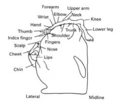
Figure 5 – the somatosensory cortex
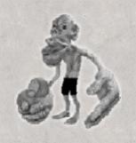
Figure 6 – the homunculus
Human skin contains several different
sense receptors that respond to mechanical and thermal
stimuli (e.g., touch, pressure, pain, cold, heat).
A sense receptor is a specialized cell
that converts a physical or chemical stimulus into action
potentials. These action potentials produced by the
receptors are conducted to the spinal cord and brain
(CNS) for processing and interpretation. The message
that is sent to the central nervous system (CNS) is
always a train of action potentials, regardless of the
kind of stimulus that excites a particular receptor.
Sensory receptors that respond to touch
send action potentials through axons that enter the
dorsal columns of the spinal cord and ascend to the
medulla of the brainstem. These axons then make connections
(synapses) with another pathway within the medulla.
It is here that the pathway crosses over the brain midline
and then continues to the thalamus. The final pathway
begins in the thalamus and continues to the specific
region of the sensory cortex.
Interesting facts:
-
When an area of the body is missing
the somatosensory cortex can reconfigure itself.
-
Homunculus means “little person”
-
Homunculus is different in different
animals
Something to think about
– We will return to this at the vision and olfaction
stations, but it is interesting to think about the fact
that we are normally not aware of our clothes touching
us, or of the seat we are sitting on. This is because
sensory receptors stop responding after an extended
period of constant stimulation, but once we move even
slightly we notice the change.
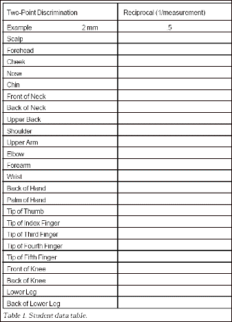
Procedure:
- What parts of the body seem more sensitive to
you? Where do you think you will have the most touch
receptors?
- Each group should choose a subject, a measurer,
and a recorder.
- Use two toothpicks, start far apart, go closer
and closer (making sure to touch the toothpicks
at the same time), asking the subject if they feel
two points or one (they’re blindfolded/closing
their eyes), continue until they don’t feel
two. (Touch randomly with one to keep them on their
toes) Record when they can’t feel two points
on each of the different body regions in the Table
1 (next page).
- Calculate the reciprocals (1/measurement)—the
bigger the reciprocal, the more touch receptors
and the larger the representation on the somatosensory
cortex map.
- Make a graph of the reciprocals (or the distances).
Discussion Questions:
How does this map affect our life?
How is it evidenced in every day life?
Why is this arrangement beneficial for
us?
Visual System Station
Objectives:
- Become familiar with visual neuroanatomy
- Discover the mechanisms behind different visual
illusions and visual fatigue
Background:
The eye is derived from neural tissue,
and actually processes information (as opposed to just
transmitting it). Light enters the eye, hits the retina,
activates a series of rods and cones, which become excited
and cause retinal ganglion cells to fire. Rods and cones
differ in the wavelength of light they are most sensitive
to, and how strongly they respond to light energy.
Rods are smaller than
cones and concentrated in the periphery of the retina.
Cones are concentrated
in the fovea (center of the retina), and contain 3 different
pigments.
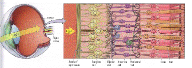
Figure 7 – the eye
Part 1: Visual system anatomy
Equipment:
-
Forceps
-
Dissected sheep brain
-
Gloves
Procedure:
- Take a look at the dissected brain in front of
you and the diagram of the visual system. What do
notice?
- Find the optic chiasm. Can you think of any reasons
why visual information would only partially cross
between hemispheres?
- Look at the diagram of the visual system (Figure
8). Find the two different pathways. Now try and
locate these structures in the dissected sheep brain.
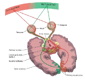
Figure 8 –
the visual system
-
Knowing that the brainstem (colliculus,
pulvinar nucleus) is a more evolutionarily primitive
structure compared to the cortex, what kinds of
visual information do you think each of these pathways
processes?
-
Why do you think the visual cortex
is broken down into so many different areas? (Figure
9)

Figure 9 – the visual cortex
Part 2: The blind spot
Introduction:
Whether you know it or not, you have a
blind spot on the retina of each eye where there no
photoreceptors.
Equipment:
- Pencil and paper
- Ruler/tape measure
Procedure:
- Make a tester by marking + on the far right side
of a piece of notebook paper.
- Stand with your back to a wall, with your head
touching the wall.
- Hold the tester 500 mm (0.5 m or 50 cm) in front
of your eye. (It may help to have someone help you.)
- Close your right eye and look at the + with your
left eye.
- Place a pencil eraser on the far left side of
the tester, and slowly move the pencil eraser to
the right.
- When the eraser disappears, mark this location
on the tester. Call this point "A."
- Continue moving the eraser to the right until
it reappears. Mark this location on the tester.
Call this point "B."
- Repeat the measurements until you are confident
that they are accurate.
- Measure the distance between the spots where the
eraser disappeared and reappeared.
- To calculate the width of your blind spot on your
retina, let's assume that 1) the back of your eye
is flat and 2) the distance from the lens of your
eye to the retina is 17 mm. We will ignore the distance
from the cornea to the lens.
- With the simple geometry of similar triangles,
we can calculate the size of the blind spot because
triangle ABC is similar to triangle CDE. So, the
proportions of the lines will be similar.
Figure 10 – the blind spot

Discussion Questions:
Why do you think you have a blind spot?
What are some ways your brain and visual
system compensates for this blind spot?
What would a larger or smaller blind spot
mean for your vision?
Part 3: Color Vision
Introduction:
As mentioned before, there are two types
of photo receptors located in the eye, rods and cones.
They respond to different kinds of light. Each rod or
cone responds to two wavelengths of light (for example,
rods respond to either red or green light). The purpose
of this next exercise is to investigate the characteristics
of rods and cones with respect to color.
Procedure:
-
-
Follow the directions on screen;
play with different colors and saturation levels.
Discussion Questions:
Why do you think you see colors that aren’t
actually there? Using what you know about how the nervous
system works, how can you explain this fatigue? Do you
think the fatigue observed visually can be applied to
other parts of the nervous system, not just sensory
systems?
Olfactory Fatigue Station
Equipment:
2 bottles of different aromatherapy scents
Stopwatch
Ruler
Notebook paper
Objectives:
Explore mechanisms behind olfactory sensation
and olfactory fatigue
Background:
Olfaction is the sense of smell. To learn
how it works, let’s imagine a certain smell in
the air. These chemical molecules enter your nasal passage
and then get trapped in the mucus layer in your nose.
Olfactory epithelium neurons project cilia (which act
as dendrites) into the mucus layer in the nasal passage
so passing molecules will bind to the receptors on the
ends of the cilia. There are hundreds of different types
of receptors which recognize thousands of smells. The
chemical molecules of each odor have a receptor that
it will fit into, like a lock and key. These neurons
have axons that lead to the olfactory bulb which is
right under the frontal lobe in the brain. Some neurons
of the olfactory bulb lead to the olfactory tubercle
where the message is continued to the limbic system,
thalamus and cortex. The thalamus and cortex are thought
to give us a conscious sense of smell while the limbic
system, which includes the amygdala and hippocampus,
is thought to be involved in the emotional experience
of smell such as inducing memories. Other neurons of
the olfactory bulb lead to the olfactory cortex where
odors can be identified.
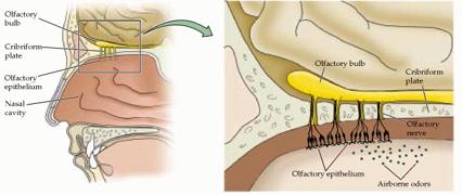
Figure 11 - Olfaction from the nose to the brain
Olfactory fatigue is when, after smelling
one specific smell for a large amount of time, the smell
is no longer noticeable. This is because the neurons
of the olfaction system become accustomed to the continual
odor molecules and stop causing action potentials (this
concept will also be discussed at the somatosensory
and visual stations). This is due to a block of ions
flowing in the receiving neuron which stops signaling
to the brain. When a new odor enters the mucus membrane,
the neurons are reactivated.

Figure 12 - An olfactory
neuron
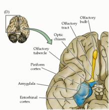
Figure13 - Olfaction in
the brain
Procedure:
- Obtain 2 bottles of aromatherapy scents.
- Have one person control the stopwatch. The other
person should be holding the scent bottle about
30 cm in front of their face.
- Press and hold your left nostril closed.
- Open the bottle and start wafting (demonstrated
by teacher) the scent toward your face and begin
the timer.
- Continue smelling until you can no longer notice
the odor and stop the timer at this point.
- Switch bottles and repeat steps 3-5 for the second
scent.
- When complete, open your left nostril and waft
the second scent under the left nostril and record
observations. What happens here and why?
- Also record any memories the scents may bring
to mind.
- Did the different smells take a different amount
of time to go away
Discussion Questions:
Why would remembering odors be important
for survival?
Why would you want to have olfactory fatigue?
What would your world be like if you lost
your sense of smell?
Corpus Callosum and Hemispheric
Communication Station
Objectives:
Background:
The left brain is primarily responsible
for language production and processing (i.e. reading,
writing, speaking), and the right brain is primarily
responsible for object/face recognition. In the case
of a normal individual with an intact corpus callosum,
information about visual stimuli can be shared between
the hemispheres through the corpus callosum even though
after passing through the optic chiasm each hemisphere
is only receiving input about one half of the visual
field. With a split-brain patient, information coming
into the right hemisphere is confined and cannot be
shared with the left, nor can the left hemisphere share
information with the right. To demonstrate, take the
below image of a male/female face:

The right half of the visual field is
a man’s face and the left is a woman’s face.
If the split-brain patient focuses on the dot, the man’s
face will project to the left hemisphere and the woman’s
face will project to the right hemisphere. When shown
a picture of a whole man’s face and a whole woman’s
face and asked to point to the image just seen, the
patient will point to the woman’s face. However,
if asked to verbally say which face was just seen, the
patient will say a man’s face. This is because
the man’s face projected to the language side
of the brain (LH) and the woman’s face projected
to the object recognition side of the brain (RH).
With normal individuals, the connectedness
of the two hemispheres can be highlighted by attempting
bimanual tasks. Since information is shared between
the hemispheres, it is nearly impossible to complete
two tasks simultaneously without there being interference
between the hemispheres (see protocol for goal b). This
activity not only focuses on the importance of the corpus
callosum in allowing information to move freely throughout
the brain, but it also speaks to the degree of lateralization
within the brain. While lobectomy patients speak to
the re-wiring capability of the brain, there are certain
basic functions that are specific to each hemisphere
of the brain (i.e. language and object recognition).
Procedure:
-
Look at the brain section at this
station, can you locate the corpus callosum?
-
Do you remember its function?
-
How does its location makes it optimal
for transferring information from one side of the
brain to the other?
-
Discuss its composition (i.e. white
matter, axons with cell bodies in either hemisphere,
etc)
-
Describe the basic functions of
each hemisphere, what kinds of things does the corpus
callosum allows us to do?
-
Discuss what this “highway”
reveals about the specificity of function in each
hemisphere
Part I - Bimanual Coordination
Procedure:
-
Try to separately draw a star with
one hand and a triangle with their other hand. How
easy is this task is when you are only concentrating
on one image and one hand?
-
Shut your eyes (or blind-fold) and
try drawing the star and triangle at the same time
now
-
Most students will make two drawings
that look very similar because the motor connections
are shared (by way of what structure? Corpus
callosum!!) between the hemispheres, making
it difficult to draw two different shapes
-
If that structure were severed so
that it could not connect the hemispheres (as in
a split-brain patient), a bimanual drawing task
would be easy
Part II - Split Brain Demo on Computer
Procedure:
-
Complete computer activity (http://nobelprize.org/medicine/educational/split-brain/index.html)
where you can guess what a split-brain patient can
or cannot process depending on which hemisphere
a series of tasks are flashed
-
Discuss your predictions with the
group
Discussion Questions:
Why do you think the brain evolved to
have hemispheric specification of function?
How would being a split-brain patient
affect our daily lives? Would it?
Can you think of any ways that split-brain
patients might learn to compensate for their hemispheric
disconnectedness?
Can you think of any sicknesses that might
require the severing of the corpus callosum?
References:
Purves, D., Augustine, G.J., Fitzpatrick,
D., Katz, L.C., LaMantia, A., McNamara, J.O., and Williams,
S.M. (2001). Neuroscience
2nd ed. Sunderland, MA: Sinauer
Associates, Inc.
http://www.nabt.org/sup/publications/nlca/toc.htm.
www.stjude.org/glossary
science.education.nih.gov/supplements/nih2/addiction/other/glossary/glossary2.htm
www.ninds.nih.gov/health_and_medical/pubs/sci_report.htm
www.addictionstudies.org/glossary_n.html
www.alz.org/Resources/Glossary.asp
www.als.net/als101/glossary.asp
science.education.nih.gov/supplements/nih2/addiction/other/glossary/glossary.htm
www.alz.org/Resources/Glossary.asp
www.nationalmssociety.org/S%20-%20Z.asp
en.wikipedia.org/wiki/Grey_matter
www.azspinabifida.org/gloss.html
en.wikipedia.org/wiki/Cerebral_hemisphere
www.theuniversityhospital.com/epilepsy/html/aboutepilepsy/glossary.htm
www.seniormag.com/conditions/cancer/cancerglossary/t.htm
webanatomy.net/anatomy/brain_notes.htm
www.fmrib.ox.ac.uk/~stuart/thesis/chapter_3/image3_10.gif
www.nabt.org/sup/publications/nlca/toc.htm
faculty.washington.edu/chudler/blindspot.html
www.nabt.org/sup/publications/nlca/toc.htm |