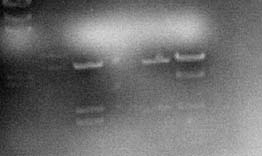
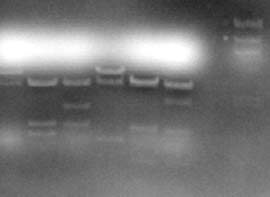
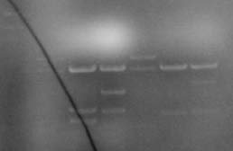
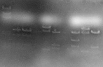
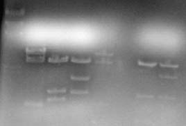
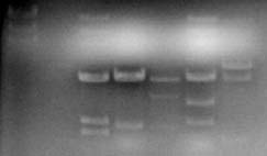
Table 1. Agarose gels containing DNA stained with DNA safe stain after electrophoresis. DNA samples were cut with restriction enzymes prior to running gel. In most cases each lane contained the following, lane 1: molecular size marker; lane 2, crime scene DNA; lane 3 suspect #1 DNA; lane 4 suspect #2 DNA; lane 5 suspect #3 DNA; lane 6 suspect #4 DNA; lane 7 suspect #5 DNA.
Gel images were captured as TIFF files in black and white (not the blue color you saw in the lab) and then imported into Adobe Photoshop where they were cropped and the brightness and contrast adjusted to optimize the quality of the images. Note that the DNA appears white in these gels. There was a lot of background dye still in the gel at the time the photographs were taken especially the bright band near the top of each gel which came from some of the running dye.
|