Remote Ready Biology Learning Activities has 50 remote-ready activities, which work for either your classroom or remote teaching.
Serendip is an independent site partnering with faculty at multiple colleges and universities around the world. Happy exploring!

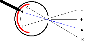
The sheet of photoreceptors is much like a sheet of film at the back of a camera. But it has a hole in it.
At one location, called the optic nerve head, processes of neurons collect together and pass as a bundle through the photoreceptor sheet to form the optic nerve (the thick black line extending up and to the left in the diagram), which carries information from the eye to the rest of the brain. At this location, there are no photoreceptors, and hence the brain gets no information from the eye about this particular part of the picture of the world. Because of this, you should have a "blind spot" (actually two, one for each eye), a place pretty much in the middle of what you can see where you can't see.

Close your left eye and stare at the cross mark in the diagram with your right eye. Off to the right you should be able to see the spot. Don't LOOK at it; just notice that it is there off to the right (if its not, move farther away from the computer screen; you should be able to see the dot if you're a couple of feet away). Now slowly move toward the computer screen. Keep looking at the cross mark while you move. At a particular distance (probably a foot or so), the spot will disappear (it will reappear again if you move even closer). The spot disappears because it falls on the optic nerve head, the hole in the photoreceptor sheet.
So, as you can see, you have a pretty big blind spot, at least as big as the spot in the diagram. What's particularly interesting though is that you don't SEE it. When the spot disappears you still don't SEE a hole. What you see instead is a continuous white field (remember not to LOOK at it; if you do you'll see the spot instead). What you see is something the brain is making up, since the eye isn't actually telling the brain anything at all about that particular part of the picture.
Alright, you say, that's kind of neat, but maybe the brain isn't "making it up." It just knows to put white where the blind spot is. Let's try another situation and see what happens.
Table of Contents: Blindspot Home Page |
Resources Elsewhere:
MD Support, a website serving the Macular Degeneration Community, adapted this exhibit for diagramming vision: http://www.mdsupport.org/map/map.html.
| Brain and Behavior Forum | Brain and Behavior | Serendip Home | |