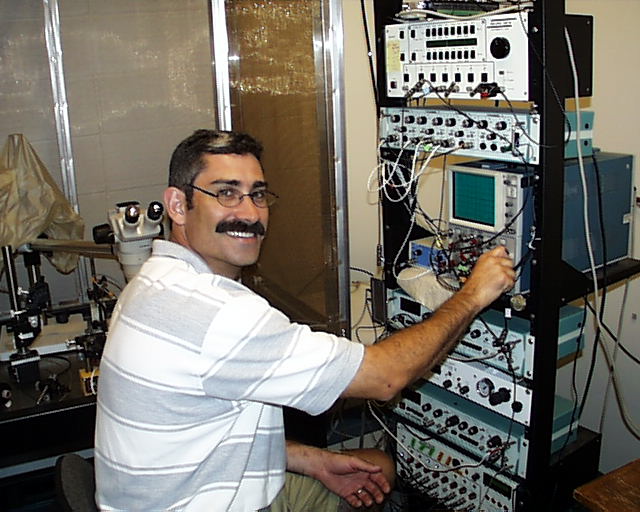
Peter D. Brodfuehrer
Associate Professor of Biology and Chair

Research in my laboratory focuses on elucidating the neuronal basis of leech swimming. For decades researchers have approached the problem of understanding the neuronal basis of animal behavior in both invertebrate and vertebrate systems by 'circuit breaking', identifying individual neurons and assessing each neuron's role in behavior. The success of this approach is clear from the number of neuronal networks that have been described that explain, to varying degrees, how motor patterns are generated and modulated at the level of synaptic interactions between identified neurons.
The neuronal basis of leech swimming is well understood at the level of synaptic interactions within and between five functional classes of neurons (mechanossensory, trigger, gating, oscillatory and motor). However, very little is known about the pharmacology and cellular events underlying most of these synaptic interactions. My laboratory has recently found that non-NMDA receptors mediate fast excitatory postsynaptic potentials (EPSPs) in the monosynaptic connections from mechanosensory P cells to trigger neurons, from trigger neurons to gating neurons, and from gating neurons to oscillatory neurons. Activation of non-NMDA receptors by trigger neurons (cells Tr1) also gives rise to a slower, depolarizing-like potentials (DPPs) in swim-gating neurons, cells 204. At low stimulation rates (< 2 Hz), cell Tr1 produces constancy latency EPSPs in cell 204, while at stimulation frequencies greater than approximately 25, cell Tr1 stimulation produces a gradual decrease in the membrane potential (3-5 mV) and an increase in the firing frequency of cell 204, which lasts for several seconds and often leads to swimming. The DPPs in cells 204 are important as they provide a mechanism for transforming a short-lasting stimulus into a long-lasting drive to the swim motor program, such as occurs following body wall stimulation in the leech.
My laboratory is currently testing the hypothesis that Tr1 stimulation activates a 'swim-activating network' in the subesophageal ganglion that produces DPPs in cell 204. Support for this hypothesis stems from recent observations that following Tr1 stimulation elevated levels of descending activity in the lateral connectives coincide with the duration of DPPs in Retzius cells. (Tr1 evoked response in Retzius cells mimics, both physiologically and pharmacologically, that in cell 204. Brodfuehrer and Friesen, 1986 and Thorogood and Brodfuehrer, 1995)
 | Correlation between descending connective activity and Retzius cell activity following Tr1 stimulation. Intracellular stimulation of cell Tr1 (top trace) depolarizes the Retzius cell (middle trace) by approx. 3-5 mV which lasts greater than 10s. Similarly, activity in a lateral connective (bottom trace) increases following Tr1 stimulation and remains elevated as long at the Retzius cell is depolarized. Cell Tr1 is located in the subesophageal ganglion; Retzius in segmental ganglion 1. Extracellular recording from the connectives between segmental ganglion 3 and 4. Vertical scale bar pertains to Retzius membrane potential only. |
Another aspect of the initiation of swimming that is not adequately explained by the known synaptic interactions comprising the swim-initiating network or pathway is the variability with which stimulating this pathway leads to swimming. For example, the probability of eliciting swimming by intracellular depolarization of cell Tr1 is highly variable both from preparation to preparation and from one stimulus trial to the next within a given preparation. Stimulation of cell Tr1 can lead to swimming on one trial, but might not on the next trial even though the strength of cell Tr1 stimulation is constant in both trials. This and other observations suggest that the 'state' of the ongoing activity in the ventral nerve cord strongly influences whether inputs lead to swimming, while a recent computational study has also shown that several time-resolved computational techniques are useful in detecting and characterizing neuronal activity in the leech nervous system that leads to swimming (Cellucci et al., 2000).
Brodfuehrer, P.D. and Thorogood, M.S (2001) Identified Neurons and Leech Swimming Behavior. Progress in Neurobiology 63:371-381.
Cellucci, C.J., Brodfuehrer, P.D., Acera-Pozzi, R., Dobrovolny, H., Engler, E., Thompson, R., Los, J. and Albano, A.M. (2000) Linear and nonlinear measures predict swimming in the leech. Physics Review E 62: 4826-4834.
Cacciatore, T.W., Brodfuehrer, P.D., Gonzalez, J.E., Adams, S.R., Tsien, R.Y., Kristan, W.B.Jr., and Kleinfeld, D. (1999) Identification of neural circuits by imaging coherent changes in membrane potential with FRET-based dyes. Neuron 23:449-459.
Thorogood, M.S., Almeida, V.W. and Brodfuehrer, P.D. (1999) Localization of glutamate receptor 5/6/7 - like and glutamate transporter-like immunooreactivity in the leech central nervous system. J. Comp. Neurol. 405:334-344.
Brodfuehrer, P.D., Debski, E.A., O'Gara, B.A. and Friesen, W.O. (1995) Neuronal control of swimming. J. Neurobio. 27:403-418.
Thorogood, M.S. and Brodfuehrer, P.D. (1995) The role of glutamate in swim initiation in the leech. Invert. Neurosci. 1:223-233.
Brodfuehrer, P.D., Parker, H.J., Burns, A. and Berg, M. (1995) Control of the segmental swim generating mechanism by an identified pair of interneurons in the leech head ganglion. J. Neurophysiol. 73:983-992.
Brodfuehrer, P.D. and Burns, A. (1995) Factors influencing the decision to swim in the medicinal leech. Neuro. of Learning and Mem. 63:192-199.