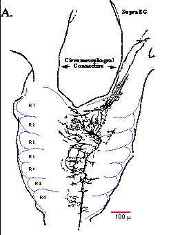
 Fig.1A. Camera lucida drawing of a Lucifer Yellow fill of cell SE1. The soma of cell SE1 lies on the dorsal aspect of the anterior division of R2 in the subesophageal ganglion. The axon of cell SE1 projects posteriorly and exits the subesophageal ganglion in the ipsilateral connective (ipsilateral with respect to the position of cell SE1's cell body). Branches of cell SE1 extend primarily along the midline of the subesophageal ganglion, with a few branches projecting anteriorly into the circumesophageal connective and supraesophageal ganglion (SupraEG; partialy drawn). Dashed lines indicate the approximate borders of the cell packets (labeled R1-R4) in the subesophageal ganglion.
Return to Results
Fig.1A. Camera lucida drawing of a Lucifer Yellow fill of cell SE1. The soma of cell SE1 lies on the dorsal aspect of the anterior division of R2 in the subesophageal ganglion. The axon of cell SE1 projects posteriorly and exits the subesophageal ganglion in the ipsilateral connective (ipsilateral with respect to the position of cell SE1's cell body). Branches of cell SE1 extend primarily along the midline of the subesophageal ganglion, with a few branches projecting anteriorly into the circumesophageal connective and supraesophageal ganglion (SupraEG; partialy drawn). Dashed lines indicate the approximate borders of the cell packets (labeled R1-R4) in the subesophageal ganglion.
Return to Results