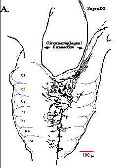
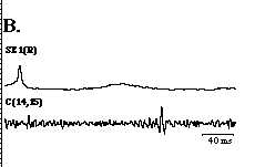
Morphology of cell SE1. A. Camera lucida drawing of a Lucifer Yellow fill of cell SE1. The soma of cell SE1 lies on the dorsal aspect of the anterior division of R2 in the subesophageal ganglion. The axon of cell SE1 projects posteriorly and exits the subesophageal ganglion in the ipsilateral connective (ipsilateral with respect to the position of cell SE1's cell body). Branches of cell SE1 extend primarily along the midline of the subesophageal ganglion, with a few branches projecting anteriorly into the circumesophageal connective and supraesophageal ganglion (SupraEG; partially drawn). Dashed lines indicate the approximate borders of the cell packets (labeled R1-R4) in the subesophageal ganglion. B. Cell SE1 connective spike. Computer-averaged connective spike (30 iterations) of cell SE1 recorded extracellularly in the ipsilateral connective between M14 and M15 (bottom trace; C(14,15)). The averaging program was triggered from a spike recorded intracellularly from the soma of cell SE1 (top trace). The slight rise in the middle of the top trace was due to the periodic occurrence of a second spike in cell SE1 that was subsequently averaged out. In this and all succeeding figures, the letter in parentheses indicates the location of the neuronal cell body, either the right (R) or left (L) side of the subesophageal ganglion or segmental ganglion, while the number indicates the segmental ganglia from which the recording is made.
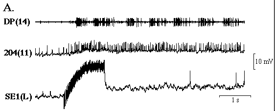
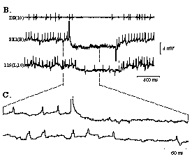
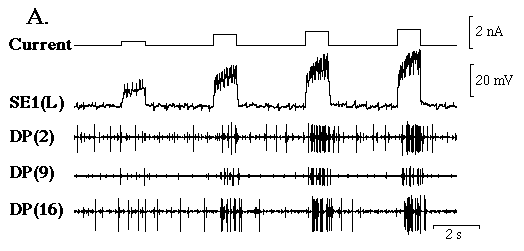
Physiological properties of cell SE1. A. Depolarization of cell SE1 (middle trace) with a short duration (1.2 s) current pulse produces a high frequency burst of action potentials (average frequency = 62 Hz) that initiates swimming. Note that membrane potential oscillations in cell 208, a swim oscillatory interneuron (bottom trace), and bursts of action potentials in the dorsal posterior (DP) of M13 begin almost immediately after intracellularly depolarizing cell SE1. Swimming in this and all subsequent figures is indicated by rhythmic bursts of actions potential in DP nerves (Kristan et al. 1974). During swimming the membrane potential of cell SE1 depolarizes 2 - 3 mV from rest (dashed line indicates the resting membrane potential of cell SE1) and its firing frequency increases from that before the onset of swimming. Membrane potential of cell SE1 returns to its resting level when swimming ends. Vertical scale bar pertains to both the middle and bottom traces.
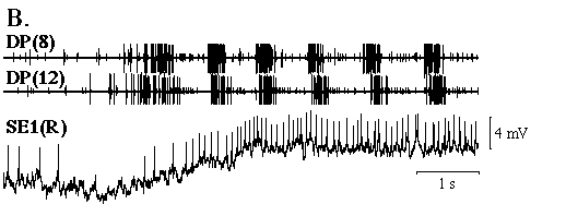
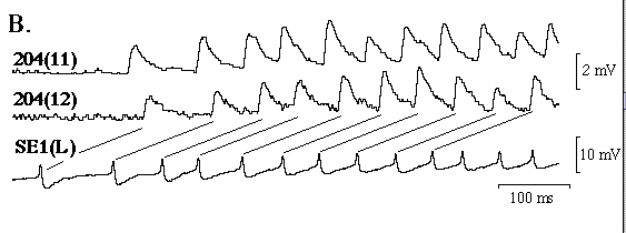
B. Spontaneous swim episode. Preceding the onset of a spontaneous swim episode, the membrane potential and spike frequency of cell SE1 increase slowly. A general increase in motor activity in DP nerves coincides with the changes in cell SE1's activity.
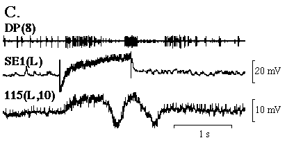
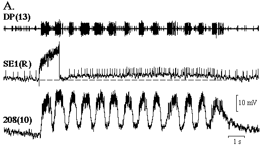
C. An abbreviated swim episode. Depolarization of cell SE1 (middle trace) rapidly excites cell 115, a swim oscillatory interneuron (bottom trace), and triggers one well-defined swim cycle in both cell 115 and DP(8) (top trace).
Stimulation of cell SE1 excites cell 204. A. Stimulation of cell SE1 (bottom trace) rapidly depolarizes and increases the firing frequency of cell 204 (middle trace). Note that swimming starts within approximately 0.5 s following stimulation of cell SE1. Vertical scale bar pertains to the bottom two traces.
B. In high Mg++ and Ca++ saline, each cell SE1 spike (bottom trace) is followed by a constant-latency EPSP in cell 204 of M11 and M12 (top two traces). The 2 mV scale bar pertains to the top and middle traces.
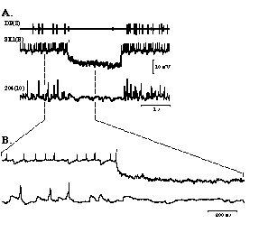
Hyperpolarizing cell SE1 (middle trace) with approximately 1 nA of current, which abolishes all spiking activity in cell SE1, eliminates all EPSPs and spiking activity in cell 204 (bottom trace), along with all large motor unit activity in DP(8) (top trace). Upon release of cell SE1 from hyperpolarization, excitatory activity immediately returns in both cell 204 and DP(8). B. Expanded section (indicated by the dashed lines) of A. In both A and B the asterisk (*) indicates the occurrence of a stimulus artifact caused by the injection of a hyperpolarizing current pulse into cell SE1. Vertical scale bar corresponds to both cell SE1 and cell 204.
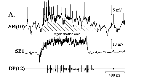
Relationship between the level of activity in cell SE1 and cell 204. A) Example demonstrating the response evoked in cell 204 (top trace) and in DP(12) by a 1 s current pulse injected into cell SE1 (middle trace). Diagonal lines indicate the parameter measured (depolarization area) tto estimate the level of exciation in cell 204.
B) Graph comparing the amount of depolarization area evoked in cell 204 as a function of cell SE1's firing frequency. In 3 preparations the amount of depolarization area induced in cell 204 increases linearly over the range of cell SE1 firing frequencies employed; r=0.92(filled squares), 0.93 (open triangles) and 0.88 (open circles).
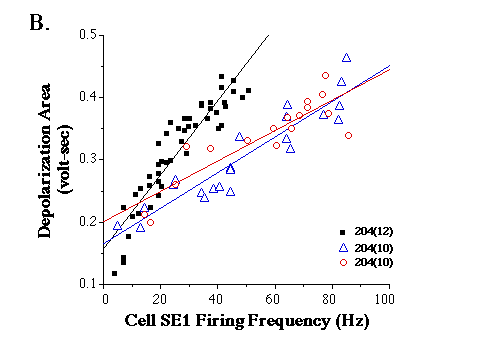
Effect of cell SE1 on the swimming rhythm. A. Phase shift. A1. Depolarization of cell SE1(R) with a 1 s current pulse during the 5 th swim cycle increases substantially the burst duration of the 5 th swim cycle recorded extracellularly in DP(10) and the period of the next swim cycle (top trace). Note that intense stimulation of cell SE1(R) produces only a slight increase in the firing frequency of its cell pair, SE1(L). Average firing frequency of cell SE1(R) during the first and second current pulses is 66 Hz and 69 Hz, respectively. A2. Graph of swim period as a function of swim cycle number. Data were calculated from swim episode in A1. Solid bar indicates the approximate timing of the second current pulse injected into cell SE1(R). B1. Period and burst duration of the swim cycles increase while cells SE1 are simultaneously depolarized. B2. Graph of swim period and burst duration as a function of swim cycle number. Data were calculated from swim episode in B1. Solid bar indicates when cells SE1 were depolarized. In A1 and B1 the vertical scale bar pertains to both the middle and bottom traces.
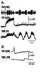
Properties of the connection from cell SE1 to cell 115. A. In high Mg++ and Ca++ saline, each cell SE1 spike (top trace) is followed by a constant-latency EPSPs in cell 115 (bottom trace). Vertical scale bar pertains to both traces. B. Hyperpolarizing cell SE1 (middle trace) with approximately 1 nA of injected current , which abolishes spiking activity in cell SE1, hyperpolarizes cell 115 and eliminates all EPSPs and a majority of the tonic spiking activity in cell 115 (bottom trace). In addition, large motor unit activity in DP(10) (top trace) ceases while cell SE1 is hyperpolarized. C. Expanded section (indicated by the dashed lines) of B. In both A and B the asterisk (*) indicates a stimulus artifact caused by the injection of a hyperpolarizing current pulse into cell SE1. In B and C the vertical scale bar corresponds to both cell SE1 and cell 115.
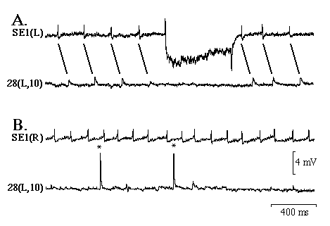 Monosynaptic connection from cell SE1 to cell 28, an oscillatory interneuron. A. In high Mg++ and Ca++ saline each cell SE(L) impulse (top trace) is followed by a constant-latency EPSP in the ipsilateral cell 28(L) (bottom trace). B. However, impulses in cell SE1(L) do not evoke a postsynaptic response in the contralateral cell 28(R). The records in A and B are from the same preparation and the experiments were performed sequentially. In B, the asterisk (*) denotes an impulse in cell 28, which is slightly distorted because the membrane potential of cell 28 has spontaneously hyperpolarized during the experiment.
 Response of cells 28 and 208 to stimulation of cell SE1. Stimulation of cell SE1 inhibits cell 28 (A.), but has no observable effect on cell 208 (B.). Note that in both A and B stimulation of cell SE1 strongly activated large motor unit activity in the DP nerve.
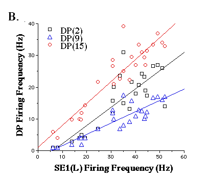
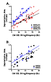 Relationship between the level of activity in cell SE1 and cell 115. A. In 3 different preparations the amount of depolarization area evoked in cell 115 increases linearly with firing frequency of cell SE1; r = 0.86(filled squares), 0.92 (open triangles) and 0.76 (open circles). B. Comparison of the effect that stimulation of one cell SE1 has on the level of excitation in the left and right cell 115. The amount of depolarization area induced in cell 115(R) and cell 115(L) in M10 by stimulation of cell SE1(R) are approximately equal at all cell SE1 firing frequencies tested; r = 0.76 (open triangles) and 0.76 (open circles).
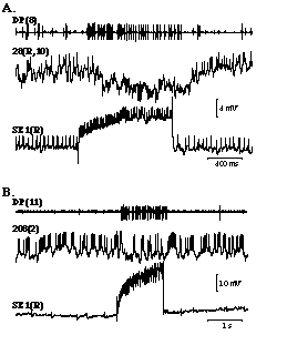
Relationship between the level of activity in cell SE1 and cell 3. A) Depolarization of cell SE1 (2nd trace) with increasing amounts of current (top trace) produces a corresponding increase in motor unit activity in all 3 DP nerves (bottom traces). The largest unit recorded extracellularly in the DP nerve is cell 3, an excitatory motor neuron that innervates the dorsal longitudinal muscles (see methods).
B) Graph showing the at the firing frequency of cell 3 recorded extracellularly from DP nerves at M2, M9 and M15 increases linearly with the firing frequency of cell SE1; r = 0.84 (filled squares), 0.89 (open triangles) and 0.92 (open circles), respectively. A and B are from different preparations.
 Cell SE1 directly excites cell 5. A. Stimulation of cell SE1(middle trace) rapidly depolarizes and increases the firing frequency of cell 5, an excitatory motor neuron to the dorsal longitudinal muscles (bottom trace), and initiates swimming (top trace). B. In high Mg++ and Ca++ saline a computer-averaged EPSP (10 iterations) is observed in cell 5 (bottom trace). The averaging program was triggered from a spike recorded intracellularly from the soma of cell SE1 (top trace). In both A and B the vertical scale bar pertains to both cell SE1 and cell 5.
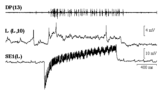
Properties of the connection from SE1 to the L-cell. A high frequency burst of action potentials in cell SE1 (bottom trace) depolarizes L-cell (middle trace) by approximately 3 mV, but elicits only 3 action potentials in the L-cell. In contrast, 39 large, motor unit action potentials occur in DP(13) in response to cell SE1 stimulation (top trace). The large, motor units are primarily action potentials from cell 3.
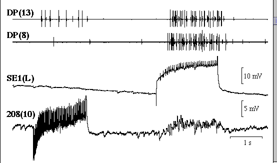
Comparison of the effects that stimulation of cell 208 and cell SE1 have on DP nerve activity, respectively. In high Mg++ and Ca++ saline, intense stimulation of cell 208 (average firing frequency = 45.5 Hz; bottom trace) elicits 7 cell 3 action potentials in DP(13) (top trace), while stimulation of cell SE1 (average firing frequency = 19.3 Hz; third trace) elicits 28 such action potentials. In addition, stimulation of cell SE1 produces summated EPSP activity in cell 208, while cell 208 stimulation has no effect on cell SE1.