
(1)
(6)
This paper reflects the research and thoughts of a student at the time the paper was written for a course at Bryn Mawr College. Like other materials on Serendip, it is not intended to be "authoritative" but rather to help others further develop their own explorations. Web links were active as of the time the paper was posted but are not updated. Contribute Thoughts | Search Serendip for Other Papers | Serendip Home Page |
Biology 202
2000
First Web Report
On Serendip
In vertebrates vision begins with light entering the pupil. The cornea and lens focus and invert the light signal and project it to the back of the eye where the retina is located. The retina consists of several layers of alternating cells and processes that convert the light signal into a neural signal, otherwise known as signal transduction. (1) Primates in particular have a fovea, a specialized area of the retina, which is composed of photoreceptors (rods and cones). The fovea allows primates to have acute vision. The optic nerve, made of ganglion cells, carries this visual information to the brain. (2)
| (1) |
(6) |
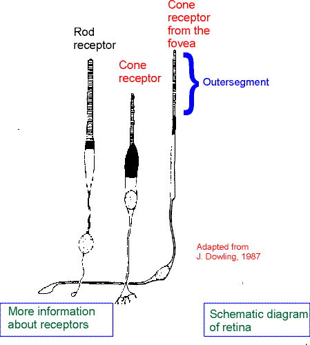
Rods and cones differ from one another in terms of their sensitive towards light and the wavelengths to which they respond. Rods, located in the periphery of the eye, are more sensitive towards light and a wider range of wavelengths when compared to cones. In the retina of primates 120 million rods can be found, however, the retina will only have approximately 6.5 million cones. (2)The rods are not color sensitive and therefore are used when there is dim light. The center of the fovea is densely populated with cones. These cones are color sensitive and "require high levels of illumination". (4)Another difference between rods and cones is that they contain different photosensitive receptors. The pigment in rods is rhodopsin, which contains opsin (protein) and retinal (the light sensitive part). Cones are also composed of a photosensitive pigment that is similar to rhodopsin. Different types of cones will contain different kinds of opsin. (2)
Opsins are extremely sensitive to light of different wavelengths and therefore this sensitivity is the foundation for color vision. (2)The eye is sensitive to light from the wavelengths of 700 nm to 400 nm; this range is known as visible light. (4)The cone cell is referred to as red (575 nm), green (535 nm), or blue (445 nm) according to what wavelength of light it is most sensitive towards and this all depends on the pigment the cone is composed of. (2)Experiments were done in 1964 to show that each human cone cell can only absorb one of those three sectors of the spectrum. (3)
Receptor cells are not the only cells that can be found in the retina. The other four cells are horizontal cells, bipolar cells, amacrine cells, and ganglion cells. The role of the receptor cells is to transduce the light signal into an electrical signal. The horizontal cells connect the rods and the cones from one region of the retina to the next. The bipolar cells connect one layer of the retina to the next requiring all information produced by the receptor cells to pass through the bipolar cells. Amacrine cells control signals from the bipolar to ganglion cells. The job of ganglion cells is to establish output from the retina to the brain as mentioned earlier. They respond to synaptic input by creating action potentials. These five cells can all give graded responses to synaptic input and amacrine cells can generate action potentials. (2)
The eye converts the light signal into a neural signal during the process of transduction. This process takes place on the outer segment of the rods and cones. George Wald discovered the first step in phototransduction in the late 1950s. He discovered the interaction a photon of light and the retinal causes the molecule to go through isomerization. This then leads to a change in the membrane potential of a rod (or cone). This hyperpolarization of any receptor cell activates the G protein known as transducin, which leads to another chain of events that trigger sodium/potassium pumps to open. Sodium is actively transported out of the cell and potassium into the cell and this causes depolarization of the membrane. The depolarization of the membrane causes an influx of calcium ions. These calcium ions cause synaptic vesicles to fuse with the presynaptic membrane of the neuron, releasing neurotransmitters into the synaptic cleft. The neurotransmitters migrate across the cleft to the postsynaptic receptors. (2)There are two types of postsynaptic receptors. One type exhibits an excitatory postsynaptic potential after receiving the neurotransmitters. This leads to the neuron depolarizing. The second kind of receptor demonstrates an inhibitory postsynaptic potential that leads to the hyperpolarization of the neuron. (6)
The rods or cones will excite the bipolar cells that are located directly underneath, which in turn excite the ganglion cells. The same process is occurring in neighboring cells. The neighboring cells excite the horizontal cells located in the retina. These cells have their processes in a laterally position which inhibit the center bipolar cell. The diffuse light excites the central bipolar cell but also inhibits it through the neighboring cells. Therefore, the ganglion cells do not become excited. However, a small spot of light will excite the bipolar cell but not the neighboring cells. Since there is no inhibition the bipolar cell will excite the ganglion cells. Another scenario is if the light only excites the neighboring cells, therefore the bipolar cells are inhibited and unable to excite the ganglion cells. These scenarios are part of the center-surrounded receptive field, which is a property of the lateral geniculate neurons. (1)
After the ganglion cells are excited and the electrical signal travels via the optic nerve to the optic chiasm. It is there that the lift and right visual worlds separate. The optic tract, fibers located after the chiasm, encircles the cerebral peduncles of the midbrain in order to reach the later geniculate nucleus (LGN). The lateral geniculate nucleus, part of the thalamus, has three layers that receive input form the left eye and another three layers that receive input from the right eye. A few of the optic tract axons branch synapse to the nuclei of the midbrain: the superior colliculi and the pretectal area. Most of the optic tract axons synapse into the LGN. (1)
Neurons in the LGN send their axons through the optic radiations to the primary visual cortex, V1. Upon reaching the V1 the axons terminate in a sub-layer of the cortex. The layers of V1 can be further subdivided into 50 layers and each of those layers divided into sub-layers. When the signal is transmitted to the upper layer of cortex, the information from the two eyes is mixed to create a binocular vision. (1)
1)Neuroscience Tutorial-Washington University School of Medicine
2)Foundations of Neurobiology by Fred Delcomyn
3)Vision, Hearing, and Smell: the Best-Known Senses
4) Color and Vision - Eastern Illinois University
6)The Joy of Visual Perception: A Web Book by Peter K.Kaiser
| Course Home Page
| Back to Brain and Behavior
| Back to Serendip |
